Ultrasound Abbreviations Meaning: How to Decode Your Scan Report


If you've just had a pregnancy scan and left feeling more confused than reassured, you're not alone.
Many women walk out of their ultrasound appointments with a report full of abbreviations, numbers, and medical terms that sound more like code than anything helpful. You might be wondering: Is everything normal? What do these measurements mean? Should I be worried?
As a registered obstetric sonographer, I’ve seen the same look of uncertainty on the faces of hundreds of clients at my clinic, Sneak-A-Peek Ultrasound, and I want to change that. This guide is here to help you make sense of your ultrasound report, so you can walk away with confidence, not confusion.
"Why are there so many abbreviations on my scan report?"
Your ultrasound report is designed for medical professionals, which is why it’s packed with shorthand. But just because it looks like another language doesn’t mean you can’t understand the basics.
Here are some of the most common ultrasound abbreviations and what they mean:
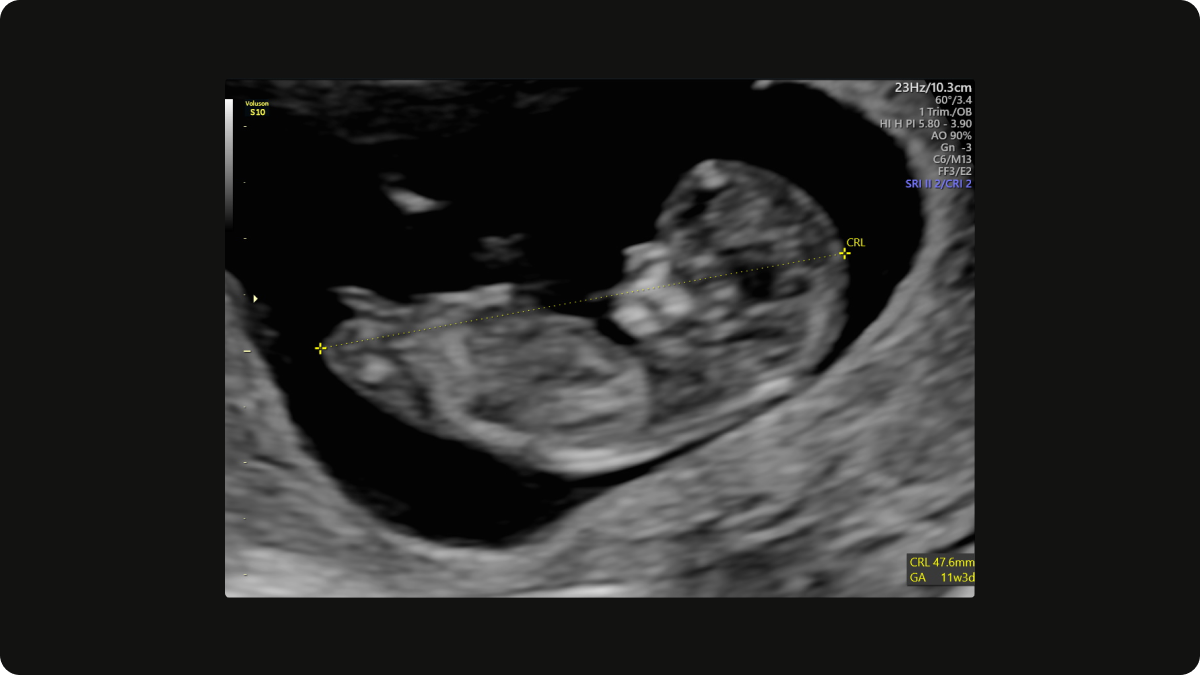
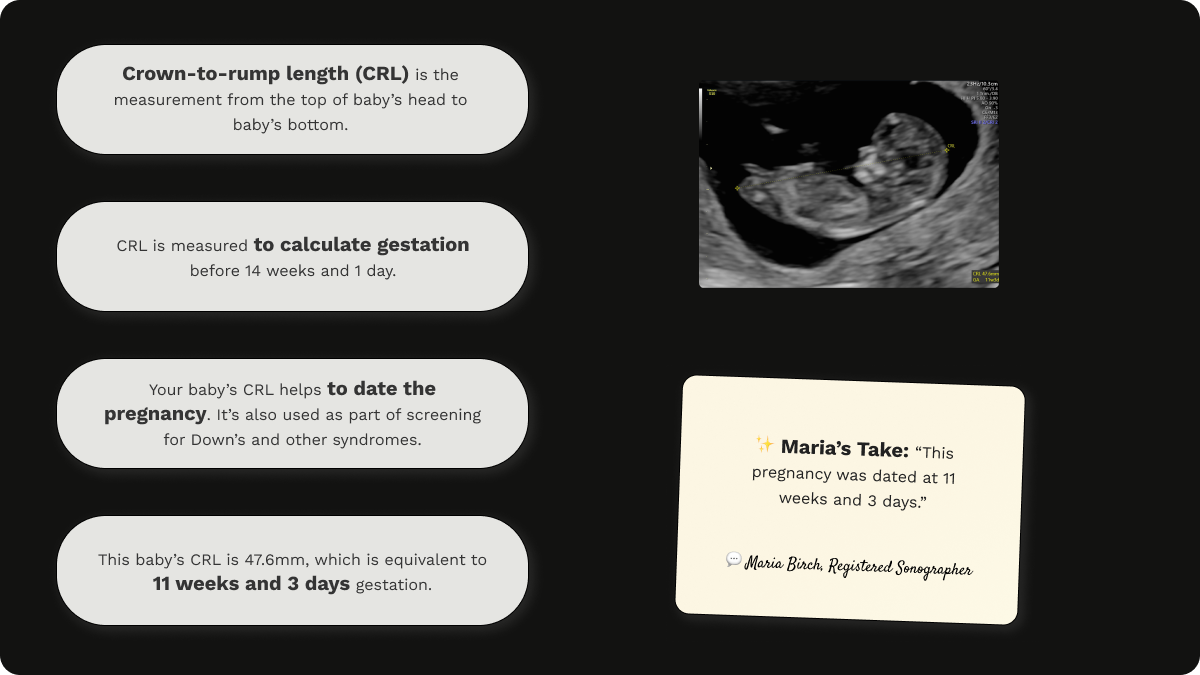
CRL (Crown-Rump Length) meaning
Crown-rump length is the measurement of your baby from head to bottom, used early in pregnancy (from 6 weeks, to 14 weeks, 1 day) to estimate how far along you are.
It is taken in the sagittal plane (from the side) with baby in a neutral position (lying flat).
It is the most accurate method of dating a pregnancy in the first trimester as it is has the least biological variability during early pregnancy, predicting the estimated due date with a margin of error +/- 3-5 days. It is more accurate than using the last menstrual period (LMP), especially in the presence of irregular cycles.
The estimated due date (EDD) determined by the CRL measurement in the first trimester becomes the 'official' estimated due date (EDD) used for the remainder of the pregnancy.
Crown-to-rump length (CRL) on an early pregnancy scan at 11 weeks gestation
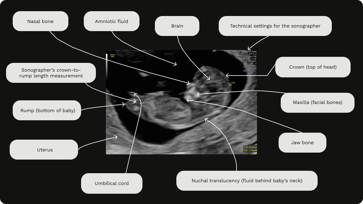
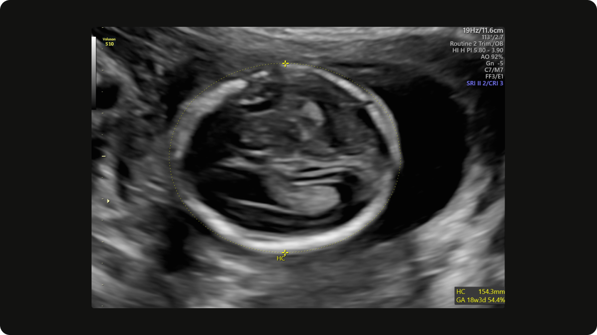
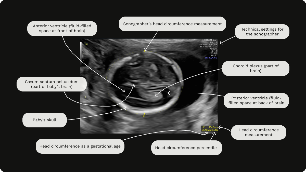
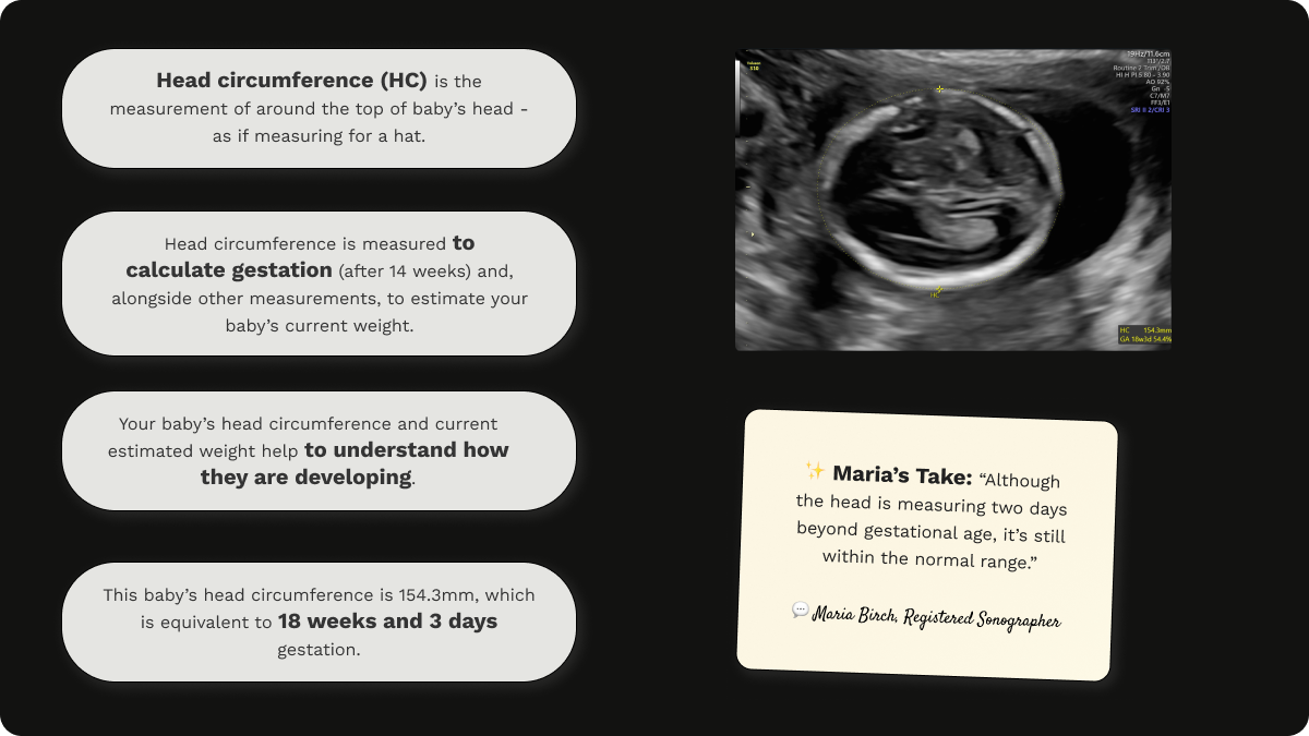
HC (Head Circumference) meaning
The head circumference is a measurement of the full distance around your baby’s head. It helps check baby's development, aids in dating a pregnancy post 14 weeks, 1 day and also contributes to the calculation of estimated fetal weight.
If your baby's head circumference is small (outside of normal range), this can be completely normal (some baby's are constitutionally small), however it can also be an indicator of fetal growth restriction (FGR). A small head circumference can also point towards certain pathologies and abnormalities, such as microcephaly (a condition related to brain development issues), certain infections such as Zika virus and chromosomal abnormalities such as Trisomy 13 (Patau's syndrome) and Trisomy 18 (Edward's syndrome).
A large head circumference (outside of normal range) can also be completely normal - some families do have big heads naturally, however some syndromes such as Sotos syndrome, can present with a larger head circumference. It can also be a sign of intracranial masses or abnormalities such as arachnoid cysts, tumours or encephaloceles.
Head circumference (HC) on a gender scan at 18 weeks gestation
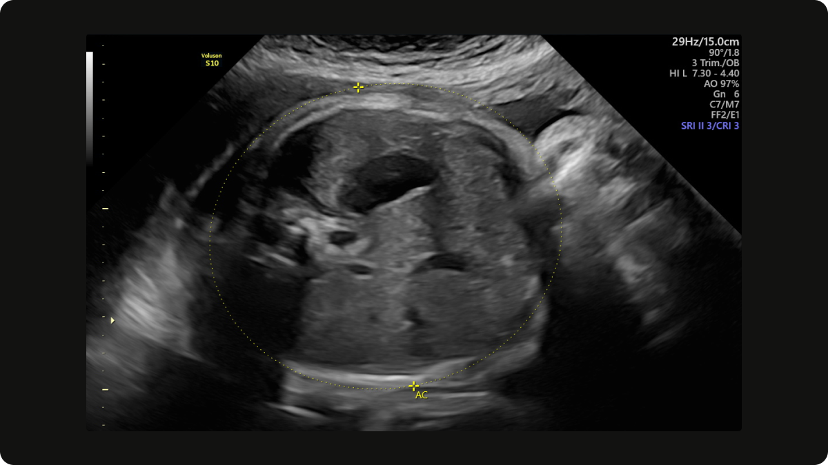
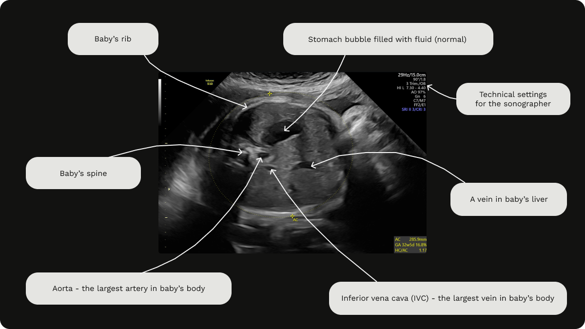
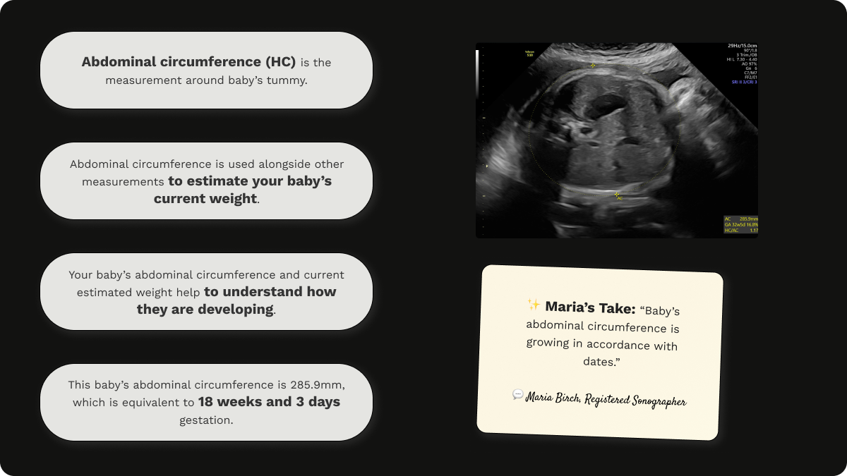
AC (Abdominal Circumference) meaning
Abdominal circumference is the measurement around your baby’s tummy. It helps check baby's development, and also contributes to the calculation of estimated fetal weight.
If your baby's abdominal circumference is small (outside of normal range), this can be an indication of fetal growth restriction (FGR) or a small for gestational age (SGA) baby.
A large abdominal circumference (outside of normal range) can be a sign of gestational diabetes, and would prompt a glucose tolerance test (GTT) for confirmation. It can also indicate a large for gestational age baby (LGA), also called fetal macrosomia. While not always a cause for concern, it may warrant further investigation and discussion with a healthcare provider as LGA babies may have an increased risk of complications during labour and delivery, such as shoulder dystocia (shoulder getting stuck).
Abdominal circumference (AC) on a growth scan at 32 weeks gestation
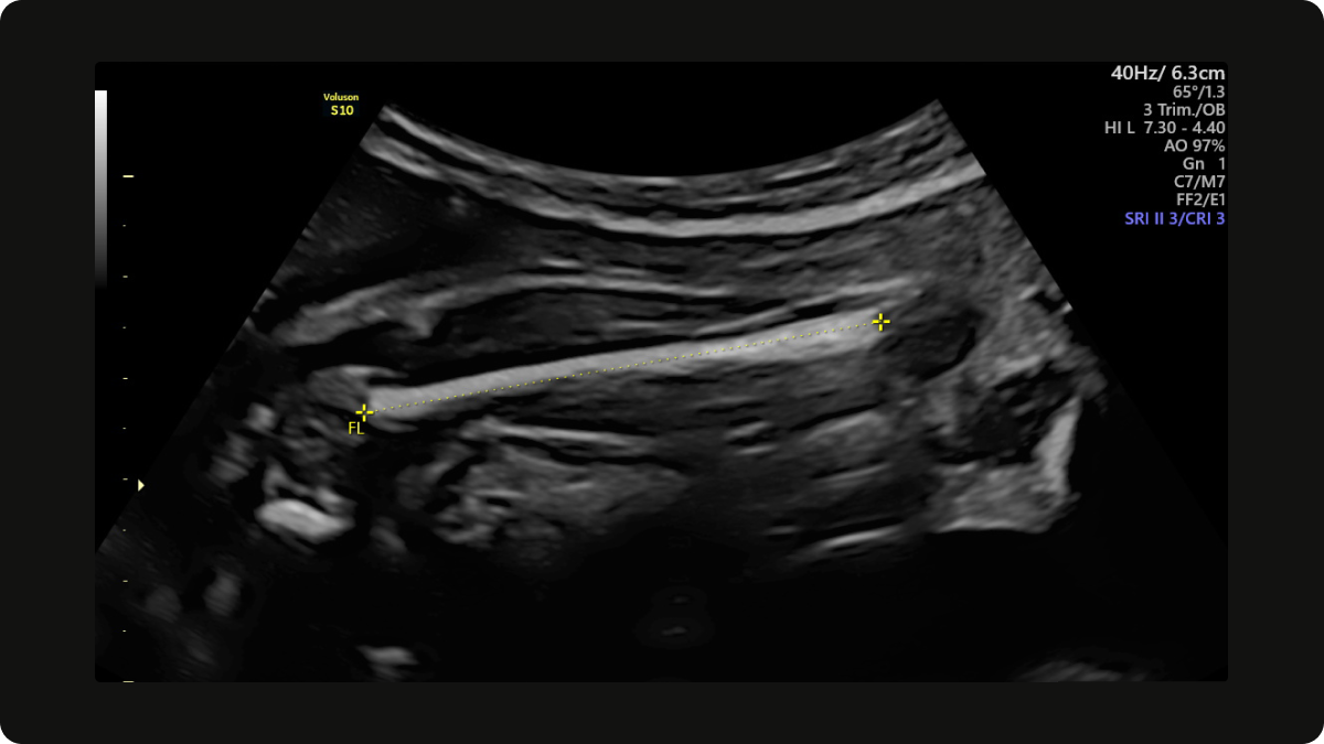
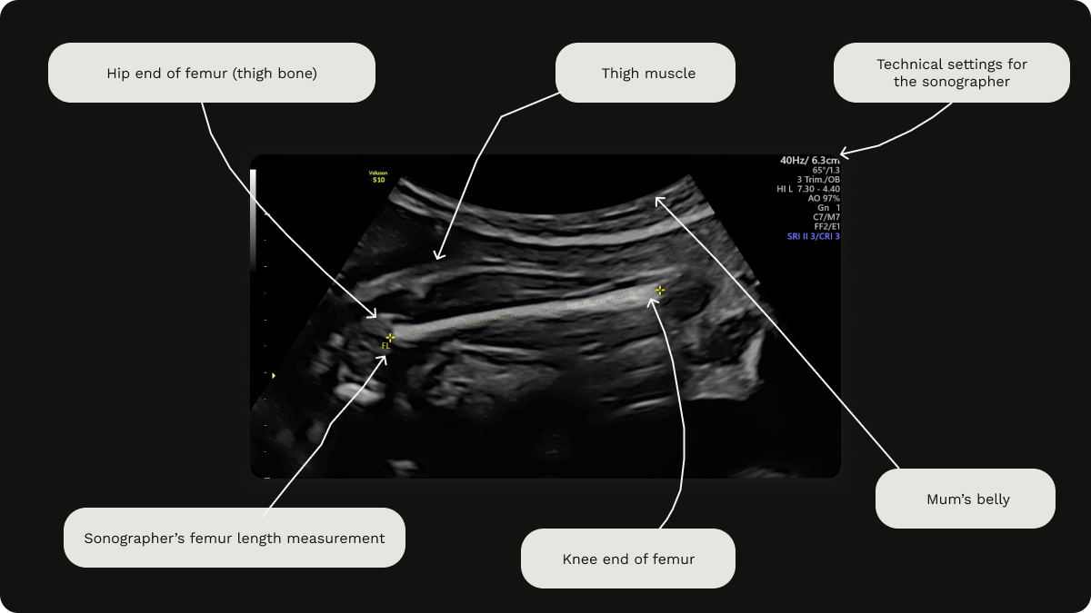
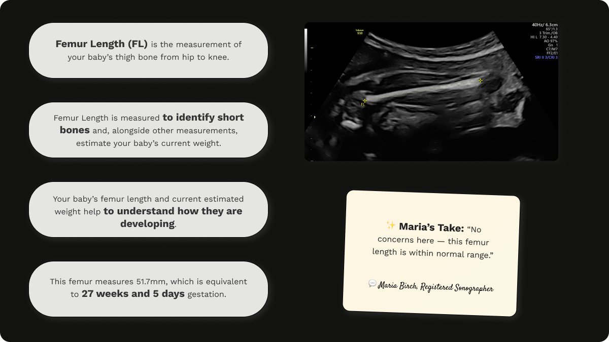
FL (Femur Length) meaning
Femur length is the length of your baby’s thigh bone. It's measured to identify short bones, but also to estimate baby's current weight.
If your baby's femur is short (outside of normal range), your sonographer will measure the length of the other femur and the length of both upper arms, shoulder to elbow. It's very important to note that an isolated short femur is likely normal, especially with petite parents.
A short femur, along with short upper arms (humerii), can be a sign of achondroplasia (dwarfism) and other skeletal abnormalities. Short femurs can also be a sign of Down's syndrome.
If a short femur is identified at your scan, further, more detailed investigation will be offered.
Femur length (FL) on a growth scan at 28 weeks gestation
EFW (Estimated Fetal Weight) meaning
Estimated Fetal Weight (EFW) is a calculated weight based on Head Circumference (HC), Abdominal Circumference (AC), and Femur Length (FL). It is not a direct measurement, but using the HC, AC, and FL, the EFW is derived by the ultrasound machine software using a formula based on biometric data.
The estimated fetal weight (EFW) is used to assess baby's growth and size from 14 weeks, 1 day.
It helps to assess baby's growth pattern by tracking a trajectory of fetal weight over time. In turn this enables the detection of small babies and large babies. This is critical in high risk pregnancies such as those with gestational diabetes or fetal growth restriction (FGR) as it allows for timing of delivery to be planned. Estimated fetal weight is especially helpful in monitoring twin pregnancies, as it facilitates the detection of discordant growth or TTTS (twin-to-twin transfusion syndrome).
As with all ultrasound measurements, EFW has it's limitations and it's accuracy can be +/- 10-15%, especially in later pregnancy. It is important to remember that it is not reliable for predicting exact birth weight. It is also highly influenced by operator skill, fetal position and maternal body habitus.
BPD (Biparietal Diameter) meaning
The width of your baby’s head, measured from one side to the other. This is no longer used in the NHS or the UK (which is why I don't have an image to share with you). BPD was replaced by HC (head circumference) because it’s less reliable and more prone to error. BPD might still appear in scan reports, but it is not used for official dating or plotting on growth charts in the UK.
The main reasons BPD fell out of favour in the UK is because:
- BPD measurement is highly effected by head shape. As it is a single line measurement from one parietal bone to the other, you can imagine that underestimation of head size would occur in longer heads, and overestimation would occur in rounder heads.
- Head Circumference (HC) captures total cranial growth, making it more reliable.
- Research has shown that HC provides better correlation with gestational age and is therefore more consistent for dating. The UK Fetal Anomaly Screening Programme (FASP) endorses HC over BPD for this reason.
Note: If you're earlier in pregnancy (before 13 weeks), your report may just include CRL and a gestational age (GA) based on that measurement.
"What do the numbers mean? Are they normal?"
Every number in your report comes with a gestational age range. For example, a CRL of 45 mm would suggest you're around 11 weeks pregnant. These values are compared to charts that reflect thousands of healthy pregnancies.
This is super helpful when it comes to dating a pregnancy. But once gestation is established in the first trimester, here’s what really matters: trends over time and whether your baby's measurements are within the expected range—not whether every single number is “perfect.”
If your scan was part of a growth check, you might also see:
- Centile or Percentile (e.g. 50th centile): This tells you how your baby's size compares to others at the same stage. 50th is average, 10th is smaller than average, 90th is bigger than average.
- AFI (Amniotic Fluid Index) or DVP (Deepest Vertical Pocket): These relate to the amount of fluid around your baby—not too little, not too much.
"What if something looks abnormal?"
First, breathe.
Ultrasound is incredibly sensitive—it can pick up tiny variations that often turn out to be normal. In many cases, a follow-up scan or further review is all that’s needed.
Common notes you might see include:
- Echogenic bowel: A brighter-than-usual area in your baby’s tummy. It often resolves on its own, however is considered as a 'soft marker' and can be associated with other abnormalities.
- Choroid plexus cyst: A small fluid-filled space in the brain, common and usually harmless.
- Short FL or small HC: May simply be a family trait, especially if both parents are petite.
If anything on your report is flagged for further review, your healthcare provider should explain what it means and next steps. But if you're still unsure, you can always ask for a second opinion.
"Can I decode my scan myself?"
You can absolutely learn to understand the basics—but always speak to a qualified sonographer or midwife before jumping to conclusions. It’s easy to spiral when you search things out of context.
At The Scan Lady, we help women every day make sense of their scan results in plain English. Whether you want to talk it through or just double-check what you've been told, we’re here to support you.
Reassurance is just one step away.
If you're still feeling unsure about your ultrasound report, don't sit with the stress. Download our free pregnancy scan timeline.